
A549 (人非小细胞肺癌细胞)(STR鉴定)
人非小细胞肺癌细胞A549来源于58岁的白人男性肺癌组织,由D.J. Giard等人建立于1972年。M. Lieber发现A549可以通过胞苷二磷酸胆碱途径合成高比例的不饱和脂肪酸卵磷脂。
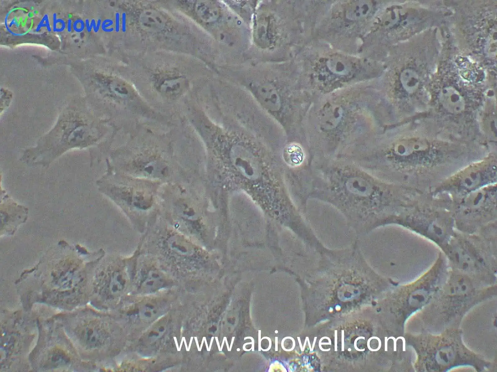
01产品图片
02产品简介
人非小细胞肺癌细胞A549来源于58岁的白人男性肺癌组织,由D.J. Giard等人建立于1972年。M. Lieber发现A549可以通过胞苷二磷酸胆碱途径合成高比例的不饱和脂肪酸卵磷脂。
03产品介绍
人非小细胞肺癌细胞A549来源于58岁的白人男性肺癌组织,由D.J. Giard等人建立于1972年。M. Lieber发现A549可以通过胞苷二磷酸胆碱途径合成高比例的不饱和脂肪酸卵磷脂。
2) 形态:上皮细胞样,多角形;贴壁生长
4) 培养条件:F-12K培养基 +10%优质胎牛血清+1%双抗 (推荐货号AW-MC004)
该细胞复苏后不耐低温和长时间运输,复苏后沿途最低温度需高于10摄氏度且运输路途不可过长。冬天常温邮寄A549细胞时会有细胞脱落、坏死等现象,请尽量选用干冰冷链快递。
1) 收到细胞后,活细胞首先观察培养瓶是否完好,培养液是否漏液,培养基是否浑浊;冻存细胞是否干冰已挥发完,冻存管盖是否脱落,破碎,若有这类情况,请务必拍照记录,并于收货24h内与我们联系。
复苏的细胞:如果是T-25培养瓶活细胞,收到后请用75%的酒精对培养瓶表面进行消毒处理,然后转入培养箱中静置2~3h后再进行后续处理。
备注:运输用的培养基不宜再次用来培养细胞,请按照 新配置完全培养基来培养细胞。
冻存细胞:如果是干冰运输的冻存细胞,收到后请立即转入液氮存储或者短暂(24h)放置-80度冰箱保存,或者直接进行细胞复苏。
认真做好每一个抗体, 每一个抗体都经过反复验证!
1) 来源:人,男性58岁, 肺部
空气,95%;二氧化碳,5%
细胞复苏、传代及冻存流程参考
1) 配制完全培养基:基础培养基+胎牛血清+双抗(特殊培养基特殊配置)
2) 细胞复苏:取5ml完全培养基于15ml离心管中,37℃水浴锅预热,从液氮管(或者-80度冰箱)中快速取出冻存的细胞,放入37℃水浴锅中,摇晃使快速化冻(1min左右),然后将化冻的细胞和预热的培养基,移入超净工作台中,化冻的细胞加入到含预热培养基的15ml离心管中,1000rpm离心5min
3) 吸弃上清,得到细胞沉淀,用2ml完全培养基轻轻重悬细胞,加入到T25培养瓶中,做好标记,放入37℃,5%CO2饱和适度培养箱中培养(培养皿复苏效果更好)
4) 24h后,观察细胞贴壁情况(未贴壁的即为死细胞--针对贴壁细胞),吸弃旧培养基,加入新鲜的预热(室温或37℃)的完全培养基,继续培养。
1) 待细胞生长到80%-90%汇合度时,吸弃旧的培养基,加入1ml无菌PBS润洗一次,以去除残余的培养基及血清(血清含有胰酶的抑制因子),然后加入1ml 0.25%胰酶,37℃培养箱中消化(1~2min左右,不同细胞消化时间不同),取出细胞,镜下观察细胞至细胞皱缩变圆
2) 加入1ml完全培养基(含FBS)终止消化,轻轻拍打,使细胞脱落下来成单个细胞悬液,收集细胞于15ml无菌离心管中,1000rpm,离心5min
3) 收集细胞沉淀,完全培养基重悬,一分为二(可根据细胞生长速度调整比例),分别加入到2个新的培养瓶中,做好标记,放入培养箱中培养。
1) 按照细胞传代方法,在超净工作台内消化收集细胞沉淀,取少量细胞用于计数
2) 用预冷的1ml冻存液(90%完全培养基+10%DMSO)或者无血清细胞冻存液重悬细胞,加入到1.2ml冻存管中,密度为1*106个/ml。
3) 放入程序冻存盒,-80℃过夜后,转入液氮长期保存。
Bevacizumab (Bev) resistance limits therapeutic efficacy in ovarian cancer (OC) patients. We identified ESM1 as a key gene in Bev-resistant OC. ESM1 secreted by OC-resistant cell lines activates the ITGB1/FAK axis to induce neovascularization and Bev resistance. Additionally, ESM1 overexpression promoted the growth and Bev resistance of OC, lung, intestinal, and hepatocellular carcinoma tumors. Then, we identified TRIM28 as an upstream regulator that stabilizes ESM1 by promoting SUMOylation, inhibiting its proteasomal degradation. In OC mice, TRIM28 overexpression promotes angiogenesis and Bev resistance via ESM1-mediated ITGB1/FAK activation. This work unveils a new molecular pathway underlying Bev resistance in OC and proposes TRIM28 and ESM1 as potential therapeutic targets.
There have been data showing that LINC01001 is highly expressed in lung cancer, but the effect of M2 macrophage exosomal LINC01001 to METTL3, glycolysis and immunity in non-small cell lung cancer (NSCLC) has not been reported. In this study, we aimed to explore the regulatory effect and mechanism of M2 macrophage exosomal LINC01001 in NSCLC. The results of our study show that the verification of macrophage exosomes, it was confirmed that exosomes regulated proliferation, glucose intake, lactate production and ATP levels of NSCLC cells. Exosomes also promoted the expression of METTL3. Bioinformatics screening showed that LINC01001 regulated METTL3. Subsequent experiments revealed exosomal LINC01001 influenced the glycolysis processes of NSCLC cells. Through RIP, it was proved that LINC01001 functioned in combination with METTL3. Bioinformatics predicted that NASP was a METTL3-targeted gene. LINC01001 could also regulate NASP methylation. Tumorigenesis in mice also indicated that LINC01001 mediated METTL3 to stimulate the development of tumors. In this study, LINC01001 was successfully verified in the exosomes-derived from M2 macrophages. It was confirmed that LINC01001 could interact with METTL3 and regulate glycolysis process in NSCLC cells. LINC01001 also inhibited T cell proliferation.
Authors:Xu Li, Li Kang, Li Jia, Xu Fang, Liang Shuzhi, Kong Yi, Chen Bolin CELLULAR AND MOLECULAR LIFE SCIENCES IF:6.2 Authors:Xuan Weixia, Wu Xu, Zheng Longcheng, Jia Huayun, Zhang Xiaoju, Zhang Xulong, Cao Bin Authors:Xujun Feng, Yuan Shi, Fang Yuan, Yanxia Hu, Xiangdong Tang, Wei Zhang, Jiadi Gan, Longhua Sun, Lingling Cao
Abnormal N6-methyladenosine (m6A) methylation in RNA plays a pivotal role in the pathogenesis of many types of tumors by influencing mRNA metabolism, alternative splicing, translocation, stability and translation. However, the specific regulators and underlying mechanisms of m6A modification in the progression of lung adenocarcinoma are not well understood. In this study, we analyzed the RNA-seq transcriptome data downloaded from The Cancer Genome Atlas (TCGA) database, and identified “m6A writer” RNA binding motif protein 15 (RBM15) expression was significantly elevated in lung adenocarcinoma (LUAD) biopsies, and the higher RBM15 levels were correlated with the poorer overall survival (OS) of LUAD patients. Further study confirmed RBM15 was prominently expressed in LUAD tissues and cell lines. Moreover, silencing RBM15 in PC9 and H1299 cells reduced cell proliferation both in vitro and in vivo , while overexpression of RBM15 in A549 cells promoted cell growth. Mechanistically, lactate dehydrogenase A (LDHA) acted as a downstream target of RBM15. RBM15-mediated m6A modification of LDHA mRNA enhanced its stability to exert an oncogenic role in LUAD. Taken together, our findings suggest that the RBM15/LDHA axis might be a novel and promising therapeutic target for LUAD.
This study aimed to elucidate the underlying mechanisms regarding microRNA-708-5p (miR-708-5p) in lung adenocarcinoma (LUAD). Here, the co-culture system of LUAD cells and macrophages, as well as a xenograft mouse model, were established. High levels of miR-708-5p were observed in LUAD. Exosomal miR-708-5p facilitated M2-like phenotype polarization, whereas miR-708-5p inhibition blocked the polarization. Exosomal miR-708-5p was identified as a pivotal signaling molecule for macrophages to mediate tumor cell proliferation, invasion, migration and IFN-γ production in T cells. In addition, miR708-5p was observed to induce PD-L1 expression, and PD-L1 silencing inhibited macrophage-induced tumor cell growth behavior and regulated CD8 T cell activity. In xenograft models, miR-708-5p inhibition and PD-L1 silencing attenuated macrophage-induced tumor growth, induced IFN-γ secretion and CD8 expression, and modulated the PTEN/AKT/mTOR pathway. In LUAD patients, there was an upregulation of both miR-708-5p and PD-L1 expression, accompanied by the activation of PTEN/AKT/mTOR. In conclusion, this study demonstrated the induction of M2 macrophage polarization and PD-L1 expression by exosomal miR-708-5p. We observed that exosomal miR-708-5p mediated the PTEN/AKT/mTOR pathway, diminished CD8 T cell activity and accelerated LUAD progression. The inhibition of specific exosomal miRNA secretion and anti-PD-L1 in the LUAD microenvironment may represent a promising avenue for LUAD immunotherapy.
Authors:Li Xu, Kang Li, Jia Li, Fang Xu, Shuzhi Liang, Yi Kong, Bolin Chen
Background Lung adenocarcinoma (LUAD) is a prevalent subtype of lung cancer (LC) whose progression is regulated by multiple genes. This study sought to find the impact and mechanism of HNRNPH1 on LUAD. Methods The expression and role of HSP90AB1, HNRNPH1, and autophagy-related protein MAP1LC3B in LUAD were detected. Additionally, bioinformatics analysis, silencing and overexpression techniques, and in vivo modeling were used to explore the regulatory mechanisms of these proteins in the progression of LUAD. Results HSP90AB1 showed high expression in LUAD and was linked to a worse prognosis. Overexpression of HSP90AB1 significantly promoted the malignant phenotype of LUAD cells and inhibited MAP1LC3B-mediated autophagy. However, overexpression of HNRNPH1 could reverse the malignant phenotype resulting from HSP90AB1 overexpression and promote MAP1LC3B-mediated autophagy by binding to HSP90AB1 mRNA and inhibiting its protein expression. Animal experiments also revealed that overexpression of HNRNPH1 could inhibit tumor progression by promoting cellular autophagy. Conclusions We verified the key role of HSP90AB1, HNRNPH1, and MAP1LC3B in LUAD, and revealed a possible regulatory mechanism, namely, HNRNPH1 could inhibit the development of LUAD by promoting autophagy through the HSP90AB1/MAP1LC3B axis. These findings may offer new insights for improving the treatment and prognosis of LUAD. Clinical trial number Not applicable.
Authors:Li Rong, Li Fen, Liu Qian, Wu Xu, Tan Xiaowu Authors:Yang Min, Chen Yanping, Huang Xueshan, Shen Fang, Meng Yanni Authors:Chen YanLi, Xu Xinyao, Zhang Jipeng, Jia Chenghui, Liang-Guan, Zhao Qirui, Jing Pengyu, Lu Qiang
Background Lung cancer exhibits high mortality and incidence rates, with tumor-associated macrophages (TAMs) serving as critical contributors to cancer progression. This study investigates the unexplored mechanistic role of HRD1—an E3 ubiquitin ligase implicated in cancer — in orchestrating TAM polarization to affect lung cancer pathogenesis. Methods HRD1 expression in lung cancer using TCGA database and validated its impact via IHC. THP-1 cells and macrophages isolated from murine tumor tissues via magnetic bead sorting were transfected with the oe-HRD1 plasmid, followed by flow cytometry, ELISA, and RT-qPCR assays to investigate HRD1's regulatory effects on macrophage polarization and function. Co-IP was employed to investigate interactions between USP7 and HRD1/PD-L1, while Immunofluorescence elucidated underlying mechanisms. Results HRD1 was highly expressed in lung cancer and promotes tumor growth in tumor-bearing mice and proliferation in THP-1 cells. Strikingly, both in vivo and in vitro overexpression of HRD1 drove macrophage M2 polarization. Mechanistically, USP7 interacted independently with HRD1 and PD-L1, while HRD1 binding to USP7 facilitated PD-L1 ubiquitination. Furthermore, HRD1 overexpression upregulated USP7 expression, thereby enhancing M2 polarization. Conclusion HRD1 promotes lung cancer progression by regulating TAM M2 polarization via USP7, offering novel therapeutic targets and diagnostic perspectives for early-stage lung cancer intervention.
Authors:Yezhou Xia, Xiaowu Tan, Saili Zeng, Yixia Jiang 细胞描述: GH3细胞系是由Tashjian AH等在1965年7月从一只7月龄的雌性Wistar-Furth大鼠的垂体肿瘤中分离建立的。GH3细胞系不是直接来源于GH1细胞系的克隆,而是从原代培养的GH1细胞在大鼠身上传代两次形成的肿瘤中建立的。上皮样的GH3细胞比GH1分泌更高水平的生长激素,也可产生细胞描述: 胶质细胞株C6是由Benda等用N-nitrosomethylurea诱导的大鼠胶质瘤克隆,并经过一系列的体外培养和动物传代交替后建成的。当细胞从低密度生长到满瓶时,S-100产量增加10倍。 细胞特性: 1)来源:大鼠,胶质瘤 2)形态:成纤维细胞样,贴壁生长 3) 6 cells 细胞描述: 克隆Neuro-2A是由R·J·Klebe和F·H·Ruddle经A株白鼠的自生肿瘤建立。该细胞产生大量微管蛋白,此蛋白在神经细胞中起应答轴浆流动的收缩系统作用。 细胞特性: 1)来源:脑神经母细胞瘤 2)形态:阿米巴样干细胞,贴壁生长 3) 6 cells 4)培养条件: MEM+
细胞描述: 此AML12(α小鼠肝脏12)细胞系由来自针对人TGFα的转基因小鼠(CD1菌株,品系MT42)的肝细胞建立。通过电子显微镜观察,这些细胞表现出典型的肝细胞特征,例如过氧化物酶体和胆小管样结构。AML12细胞保留表达血清(白蛋白,α1抗胰蛋白酶和转铁蛋白)和间隙连细胞描述: OVCAR-3细胞由T·C·Hamilton在1982年建系。取于患进行性卵巢腺癌病人的恶性腹水。NIH-OVCAR-3是研究卵巢癌药物抗性的一个合适的模型系统且由于存在激素受体,这对于激素治疗的评估或许是有用的。 细胞特性: 1)来源: 卵巢 2)形态:上皮细胞样,贴壁生长 3)规格细胞描述: 该细胞是从一名13岁的白人女性的骨肉瘤组织中分离建立的,表现为扁平形态、低饱和密度、软琼脂中的低克隆形成率和对化合物及病毒感染的敏感性。 细胞特性: 1)来源:骨 2)形态:上皮细胞样,贴壁生长 3) 6 cells 4)培养条件: MEM+10%FBS+1%NEAA+1 细胞描述: 该细胞系于1982年建立,从一位56岁白种男性的病变舌中间部分取得,该细胞具高密度颗粒状胞浆。 细胞特性: 1)来源:舌 2)形态:上皮细胞样,贴壁生长 3) 6 cells 4)培养条件: DMEM+10%FBS+1%P/S 空气,95%;二氧化碳,5% 37℃ 细胞接收后的处理: 1)收
细胞描述: HEK-293T细胞是293T(293tsA1609neo)细胞系(ATCC CRL-11268)的衍生物。细胞持续表达SV40抗原,该细胞常用于转染实验,转染效率较高。(转染一般以50%密度铺板,待细胞刚刚贴到皿壁上后(一般8-10小时),即可进行转染操作)。 细胞特性: 1)来源: 人胚胎肾脏 2)形这株细胞于1988年七月建株,组织提供者是一位非吸烟人士。
该人胰腺癌细胞PANC-1来源于一名56岁白人男性。1 U/ml 左旋天冬酰胺酶(L-asparaginase)可抑制其生长。此细胞可以在琼脂糖上生长。其倍增时间为52小时,G6PD活性处于低泳动性的B型。染色体研究表明模式数目为63,包括3个独特标记的染色体和1个小环状染色体。 以下二点证明它的恶性:(1)在软琼脂上和单层成纤维细胞上的快速生长,(2)注入无胸腺裸鼠后形成日益增长的成长型未分化癌。
相关资料下载:说明书(.pdf)(点击从官网下载)
04完整参数表
参数由厂商资料整理,采购前请以最终报价单与技术协议为准。
05荣誉与资质
该品牌暂未上传荣誉证书与资质文件。以下是平台可核验的公开信息,未上传项如实标注,不做推断。
- 营业执照未上传
- 品牌授权未上传
- 产品检测报告未上传
- 会员身份
- 收费会员
- 经营身份
- 原厂
- 成立年份
- 2016 年
- 所在地
- 湖南
- 收录条目
- 498 个
- 平台核验
- 平台代管
06品牌与供应商
公司产品涵盖抗体、细胞、培养基、质粒载体等多个领域,致力于为生命科学研究提供可靠的工具与解决方案。自创立以来,艾碧维始终秉持“求实、诚挚、勤勉、克己”的发展理念,坚持以自主研发与持续创新为核心动力。公司拥有百余人的团队,其中硕士及以上学历生物医学专业人才占比超过30%,包括博士4人,构建起一支结构合理、专业扎实的研发与服务队伍。通过设立研发部、生产部、市场与营销部、财务部等多功能部门,公司实现了从产品开发到市场推广的全流程管控。艾碧维不仅敢于质疑现状,积极为生命科学家突破技术壁垒提供支撑,更注重与客户的深度互动与合作,通过持续收集反馈、开展研发协作,提前预见并解决潜在问题。未来,公司将继续秉持“助力科研创新,让实验更简单”的使命,一步一个脚印地打造具有国际领先水平的技术研发与产业化平台,为全球生命科学领域的进步持续赋能。注意: 1.本公司产品仅供科研使用。
品牌资料由厂商公开信息与授权渠道核对整理;条目参数与价格以最终报价单为准。
07同类条目
同一子类内的条目才具备可比性。跨子类比较参数通常没有意义。
08同品牌其他条目
艾碧维 在本目录共 498 个条目,除本条外还有:
科研在线(TeleVM). A549 (人非小细胞肺癌细胞)(STR鉴定)(AW-CCH011)[EB/OL]. 产品目录, 2026-10-31. https://www.suprotek.cn/products/item/aw-cch011.html.
TeleVM. (2026, October 31). A549 (人非小细胞肺癌细胞)(STR鉴定)(AW-CCH011). 产品目录. https://www.suprotek.cn/products/item/aw-cch011.html
@misc{biocsp_item_aw_cch011,
  title        = {A549 (人非小细胞肺癌细胞)(STR鉴定)(AW-CCH011)},
  author       = {{科研在线}},
  year         = {2026},
  howpublished = {产品目录},
  note         = {更新于 2026-10-31,产品目录},
  url          = {https://www.suprotek.cn/products/item/aw-cch011.html}
}
GB/T 7714 用于中文论文与报告;APA 7 用于英文期刊;BibTeX 可直接导入 Zotero、EndNote 或 LaTeX。
许可:CC BY-NC 4.0 · 转载注明来源