
RAW264.7 (小鼠单核巨噬细胞白血病细胞)(STR鉴定)
此细胞株源自Abelson鼠科白血病病毒诱导的肿瘤。sIg-, Ia-抗原、Thy-1.2表面抗原阴性。此细胞株不分泌可检测到的病毒颗粒,XC斑点形成试验阴性。可以胞饮中性红并吞噬乳胶颗粒与酵母聚糖。可以抗体依赖性地分解绵羊红血球与肿瘤靶细胞。LPS或PPD处理2天可诱导分解红血球但对肿瘤靶细胞无作用。
01产品图片
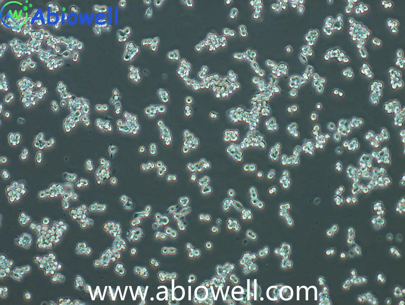
02产品简介
此细胞株源自Abelson鼠科白血病病毒诱导的肿瘤。sIg-, Ia-抗原、Thy-1.2表面抗原阴性。此细胞株不分泌可检测到的病毒颗粒,XC斑点形成试验阴性。可以胞饮中性红并吞噬乳胶颗粒与酵母聚糖。可以抗体依赖性地分解绵羊红血球与肿瘤靶细胞。LPS或PPD处理2天可诱导分解红血球但对肿瘤靶细胞无作用。
03产品介绍
此细胞株源自Abelson鼠科白血病病毒诱导的肿瘤。sIg-, Ia-抗原、Thy-1.2表面抗原阴性。此细胞株不分泌可检测到的病毒颗粒,XC斑点形成试验阴性。可以胞饮中性红并吞噬乳胶颗粒与酵母聚糖。可以抗体依赖性地分解绵羊红血球与肿瘤靶细胞。LPS或PPD处理2天可诱导分解红血球但对肿瘤靶细胞无作用。
1) 来源:鼠科白血病病毒诱导的肿瘤;单核细胞;巨噬细胞
2) 形态:不规则圆形,纺锤状,贴壁细胞,少量悬浮
4) 培养条件:DMEM+10%FBS+1%P/S (推荐货号AW-MC001)
1、细胞传代过程,无需胰酶消化,胰酶会刺激分化,直接用巴氏吸管轻轻吹打,将圆圆的细胞吹下来继续分装到新的培养瓶中即可
2、培养过程中,存在少量细胞分化,属正常现象。
3、推荐用培养皿培养,若出现难吹下来的情况,请更换培养皿培养
1) 收到细胞后,活细胞首先观察培养瓶是否完好,培养液是否漏液,培养基是否浑浊;冻存细胞是否干冰已挥发完,冻存管盖是否脱落,破碎,若有这类情况,请务必拍照记录,并于收货24h内与我们联系。
认真做好每一个抗体, 每一个抗体都经过反复验证!
空气,95%;二氧化碳,5%
复苏的细胞:如果是T-25培养瓶活细胞,收到后请用75%的酒精对培养瓶表面进行消毒处理,然后转入培养箱中静置2~3h后再进行后续处理。
备注:运输用的培养基不宜再次用来培养细胞,请按照 新配置完全培养基来培养细胞。
冻存细胞:如果是干冰运输的冻存细胞,收到后请立即转入液氮存储或者短暂(24h)放置-80度冰箱保存,或者直接进行细胞复苏。
细胞复苏、传代及冻存流程参考
1) 配制完全培养基:基础培养基+胎牛血清+双抗(特殊培养基特殊配置)
2) 细胞复苏:取5ml完全培养基于15ml离心管中,37℃水浴锅预热,从液氮管(或者-80度冰箱)中快速取出冻存的细胞,放入37℃水浴锅中,摇晃使快速化冻(1min左右),然后将化冻的细胞和预热的培养基,移入超净工作台中,化冻的细胞加入到含预热培养基的15ml离心管中,1000rpm离心5min
3) 吸弃上清,得到细胞沉淀,用2ml完全培养基轻轻重悬细胞,加入到T25培养瓶中,做好标记,放入37℃,5%CO2饱和适度培养箱中培养(培养皿复苏效果更好)
4) 24h后,观察细胞贴壁情况(未贴壁的即为死细胞--针对贴壁细胞),吸弃旧培养基,加入新鲜的预热(室温或37℃)的完全培养基,继续培养。
1) 待细胞生长到80%-90%汇合度时,吸弃旧的培养基,加入1ml无菌PBS润洗一次,以去除残余的培养基及血清(血清含有胰酶的抑制因子),然后加入1ml 0.25%胰酶,37℃培养箱中消化(1~2min左右,不同细胞消化时间不同),取出细胞,镜下观察细胞至细胞皱缩变圆
2) 加入1ml完全培养基(含FBS)终止消化,轻轻拍打,使细胞脱落下来成单个细胞悬液,收集细胞于15ml无菌离心管中,1000rpm,离心5min
3) 收集细胞沉淀,完全培养基重悬,一分为二(可根据细胞生长速度调整比例),分别加入到2个新的培养瓶中,做好标记,放入培养箱中培养。
1) 按照细胞传代方法,在超净工作台内消化收集细胞沉淀,取少量细胞用于计数
2) 用预冷的1ml冻存液(90%完全培养基+10%DMSO)或者无血清细胞冻存液重悬细胞,加入到1.2ml冻存管中,密度为1*106个/ml。
3) 放入程序冻存盒,-80℃过夜后,转入液氮长期保存。
Inflammatory bowel disease (IBD) management is often hindered by limited efficacy and significant systemic side effects of traditional oral medications, owing to their lack of targeted delivery and multifunctional capabilities. This study introduces a novel orally sequence-targeted delivery system, QT-CMOF@HAS nanocube, based on hydrophobic cross-linked CD-MOF (CMOF) for the precise delivery of quercetin (Qu) to the inflamed colonic lesions. To enhance the mitochondrial targeting capabilities of Qu, a mitochondrial-functionalized Qu-(5-carboxypentyl) (triphenyl) phosphonium bromide (TPP) precursor complex (QT) was initially synthesized and loaded into the CMOF. A chitosan/glutathione-responsive hyaluronic acid (HAS) shell was subsequently coated onto the CMOF, forming the QT-CMOF@HAS nanoplatform to further enhance the gastrointestinal tolerance. Our results demonstrated that QT-CMOF@HAS significantly alleviated colitis symptoms by modulating the repolarization of pro-inflammatory M1 macrophages to anti-inflammatory M2 phenotypes and deactivation of the TLR4/MyD88/NF-κB pathway, thus reducing the production of inflammatory cytokines. Furthermore, the integrity of the intestinal mucosal barrier and the gut microbiota were enhanced. This study highlights the potential of QT-CMOF@HAS nanocube as a promising therapeutic tool for IBD treatment, offering a multifaceted approach to tackle inflammation and support mucosal healing in a targeted and controlled manner.
Authors:Runan Zhao, Tao Chen, Shumeng Hu, Xuesong Chi, Songfeng Yu, Yanfei Li, Wenjun Wang, Beiwei Zhu, Donghong Liu, Jiangning Hu Authors:Li Bo, Li Jiayu, Zhu Zexiang, Tang Yong, Zhou Yun, Du Geshu, Li Xing ACS Applied Materials & Interfaces IF:8.3 Authors:Tao Chen, Xuesong Chi, Yangjing Li, Yanfei Li, Runan Zhao, Lihang Chen, Di Wu, Jiang-Ning Hu INTERNATIONAL JOURNAL OF BIOLOGICAL MACROMOLECULES IF:8.2
Oral ulcers are common in the oral mucosa. Frequent occurrences of oral ulcers commonly afflict patients, seriously impacting their daily life. Treatments with good anti-inflammatory and antibacterial properties are important for promoting the healing of oral ulcers. In this study, a multifunctional, soluble hyaluronic acid (HA) microneedle (MN) patch was prepared to promote oral ulcer healing. The tip layer of the MN patch was loaded with triamcinolone acetonide (TA) and epidermal growth factor (EGF) to inhibit inflammation and promote angiogenesis. Zeolitic imidazolate framework-8 (ZIF-8) was loaded onto the base layer of the MN patch, which effectively released Zn 2+ to mediate antibacterial effects. In addition, HA exerts a protective effect on the mucous membrane. Owing to these properties, the multifunctional MN patches were found to have good anti-inflammatory, antibacterial, and tissue-healing abilities, indicating that the multifunctional MN patch design successfully promoted the healing of oral ulcers.
Authors:Wenhui Ge, Yijun Gao, Liming He, Yiyu Zeng, Junhui Liu, Yi Yu, Xiaoyan Xie, Ren-ai Xu Authors:Yiyu Zeng, Yijun Gao, Liming He, Wenhui Ge, Junhui Liu, Yi Yu, Xiaoyan Xie INTERNATIONAL JOURNAL OF BIOLOGICAL MACROMOLECULES IF:7.7
Oral ulceration is the most common oral mucosal disease. Oral mucosal ulcers are extremely painful, may interfere with eating and speaking, and potentially complicate systemic symptoms in severe cases. The humid and highly dynamic environment of the oral cavity makes local drug administration for treating oral mucosal ulcers challenging. To overcome these challenges, we designed and prepared a novel dissolving microneedle (MN) patch containing multiple drugs in a core-shell to promote oral ulcer healing. The MNs contained a methacrylate gelatin shell layer of basic fibroblast growth factor (bFGF), a hyaluronic acid (HA) core loaded with dexamethasone (DXMS), and zeolite imidazoline framework-8 (ZIF-8) encapsulated in the HA-based backplane. Progressive degradation of gelatin methacryloyl (GelMA) from the tip of the MN patch in the oral mucosa resulted in sustained bFGF release at the lesion site, significantly promoting cell migration, proliferation, and angiogenesis. Moreover, the rapid release of HA and, subsequently, DXMS inhibited inflammation, and the remaining MN backing after the tip dissolved behaved as a dressing, releasing ZIF-8 for its antimicrobial effects. This novel, multifunctional, transmucosal core-shell MN patch exhibited excellent anti-inflammatory, antimicrobial, and pro-healing effects in vivo and in vitro , suggesting that it can promote oral ulcer healing.
Authors:Yi Yu, Yijun Gao, Yiyu Zeng, Wenhui Ge, Chengxuan Tang, Xiaoyan Xie, Liangle Liu
Macrophage M2 polarization plays a pivotal role in breast cancer development. The present study aimed to investigate the interplay of the Leupaxin (LPXN)/HDAC6/EGR2 axis in breast cancer and its impact on macrophage M2 polarization. Our findings indicate that LPXN overexpression in breast cancer tissues correlates with M2 macrophage polarization. To investigate LPXN's potential role, we conducted siRNA-mediated silencing in macrophages. In a breast cancer cell-macrophage co-culture system, LPXN silencing was associated with reduced cancer cell proliferation, decreased M2 polarization markers, and diminished HDAC6 expression. BIOGRD and experimental data suggest a regulatory relationship between LPXN and HDAC6. Notably, HDAC6 inhibition partially reversed the pro-M2 effects of LPXN overexpression. Further mechanistic studies revealed that HDAC6 interacts with EGR2, functioning as its deacetylase and negatively regulating EGR2 expression. EGR2 silencing partially attenuated the anti-M2 effects observed with LPXN knockdown. In murine breast cancer models, LPXN silencing was linked to increased M1 macrophage markers and reduced tumor burden. These findings suggest LPXN may influence breast cancer progression through HDAC6/EGR2-mediated regulation of macrophage polarization. In conclusion, our study demonstrated that the LPXN/HDAC6/EGR2 axis promotes breast cancer progression by augmenting macrophage M2 polarization. Graphical
Authors:He Shaozhong, Wang Qun, He Jiayi, Chen Zhongyong, Xiao Yumei COLLOIDS AND SURFACES B-BIOINTERFACES IF:5.6
To improve the osseointegration of Ti-6Al-4V implants, a Zn-, Mg-, and Cu-doped composite coating was fabricated via soft sparking micro-arc oxidation (S-MAO). The resulting S-MAO coating retained a porous microstructure and was successfully doped with the intended elements (Zn, Mg, Cu). The Zn/Mg/Cu components in the composite coating potentiated macrophage polarization towards the reparative M2 phenotype, as evidenced by an approximately 1.4-fold upregulation of CD206⁺ and a 31 % downregulation of CD86⁺. This immunomodulatory effect subsequently upregulated the expression of osteogenic genes BMP2, COL1A1, Runx2, and OCN by approximately 2.2-, 2.1-, 2.6-, and 1.7-fold, respectively. Furthermore, MC3T3-E1 cells cultured on the composite coating exhibited a characteristic spread morphology with significantly increased pseudopodia number and length, indicating enhanced cell adhesion and early osteogenic commitment. Additionally, the coating significantly enhanced ALP activity and mineralized nodule formation, which are associated with the direct osteogenic effects of Zn, Mg, and Cu. In summary, the composite coating promotes osteoblast differentiation and mineralization by modulating the immunomodulatory microenvironment and directly inducing the release of osteogenic factors. Therefore, this multi-element co-doping strategy offers a promising approach for developing bioactive titanium implants with enhanced osseointegration.
Authors:Dongying Li, Zixiong Zhou, Mengqi Li, Haimei Lin, Bo Xu, Jianfei Zhang, Zonghan Li, Xiaoming Wang, Yong Xu
This study investigated the effect of dielectric barrier discharge (DBD) on sea cucumber polysaccharide (SP-2) and evaluated its anti-inflammatory properties. The SP-2 was depolymerized by applying an input voltage of 60~90 V for 3~9 min. The features of the products were examined using high-performance gel permeation chromatography, HPLC-PAD-MS, and the Fourier transform infrared (FTIR) spectrum. The anti-inflammatory properties of the product were investigated by measuring nitric oxide (NO) release, ROS accumulation, and cell migration using RAW264.7 cells (LPS-induced or not-induced). The results showed SP-2 depolymerized into homogeneous and controllable-size oligosaccharide products. The depolymerized ratio can reach 80%. The results of the measurement of reducing sugars indicate that SP-2 was cleaved from within the sugar chain. The SP-2 was deduced to have a monosaccharide sequence of GlcN-Man-Man-Man-Man-Man based on the digested fragment information. The depolymerization product restrained the release of NO and the accumulation of ROS. By testing the RAW264.7 cell scratch assay, it was found that it enhances the migration of immune cells. DBD degradation of SP-2 leads to homogeneous and controllable-size oligosaccharide products, and this technique can be used for polysaccharide structure analysis. The depolymerized product of SP-2 has an anti-inflammatory capability in vitro.Keywords:dielectric barrier discharge;polysaccharide;degradation;RAW264.7 cell;migration
Authors:Shiwen Cheng, Han Cai, Meng Yi, Liang Dong, Jingfeng Yang
This study aimed to elucidate the underlying mechanisms regarding microRNA-708-5p (miR-708-5p) in lung adenocarcinoma (LUAD). Here, the co-culture system of LUAD cells and macrophages, as well as a xenograft mouse model, were established. High levels of miR-708-5p were observed in LUAD. Exosomal miR-708-5p facilitated M2-like phenotype polarization, whereas miR-708-5p inhibition blocked the polarization. Exosomal miR-708-5p was identified as a pivotal signaling molecule for macrophages to mediate tumor cell proliferation, invasion, migration and IFN-γ production in T cells. In addition, miR708-5p was observed to induce PD-L1 expression, and PD-L1 silencing inhibited macrophage-induced tumor cell growth behavior and regulated CD8 T cell activity. In xenograft models, miR-708-5p inhibition and PD-L1 silencing attenuated macrophage-induced tumor growth, induced IFN-γ secretion and CD8 expression, and modulated the PTEN/AKT/mTOR pathway. In LUAD patients, there was an upregulation of both miR-708-5p and PD-L1 expression, accompanied by the activation of PTEN/AKT/mTOR. In conclusion, this study demonstrated the induction of M2 macrophage polarization and PD-L1 expression by exosomal miR-708-5p. We observed that exosomal miR-708-5p mediated the PTEN/AKT/mTOR pathway, diminished CD8 T cell activity and accelerated LUAD progression. The inhibition of specific exosomal miRNA secretion and anti-PD-L1 in the LUAD microenvironment may represent a promising avenue for LUAD immunotherapy.
Authors:Li Xu, Kang Li, Jia Li, Fang Xu, Shuzhi Liang, Yi Kong, Bolin Chen 细胞描述: GH3细胞系是由Tashjian AH等在1965年7月从一只7月龄的雌性Wistar-Furth大鼠的垂体肿瘤中分离建立的。GH3细胞系不是直接来源于GH1细胞系的克隆,而是从原代培养的GH1细胞在大鼠身上传代两次形成的肿瘤中建立的。上皮样的GH3细胞比GH1分泌更高水平的生长激素,也可产生
相关资料下载:说明书(.pdf)(点击从官网下载)
04完整参数表
参数由厂商资料整理,采购前请以最终报价单与技术协议为准。
05荣誉与资质
该品牌暂未上传荣誉证书与资质文件。以下是平台可核验的公开信息,未上传项如实标注,不做推断。
- 营业执照未上传
- 品牌授权未上传
- 产品检测报告未上传
- 会员身份
- 收费会员
- 经营身份
- 原厂
- 成立年份
- 2016 年
- 所在地
- 湖南
- 收录条目
- 498 个
- 平台核验
- 平台代管
06品牌与供应商
公司产品涵盖抗体、细胞、培养基、质粒载体等多个领域,致力于为生命科学研究提供可靠的工具与解决方案。自创立以来,艾碧维始终秉持“求实、诚挚、勤勉、克己”的发展理念,坚持以自主研发与持续创新为核心动力。公司拥有百余人的团队,其中硕士及以上学历生物医学专业人才占比超过30%,包括博士4人,构建起一支结构合理、专业扎实的研发与服务队伍。通过设立研发部、生产部、市场与营销部、财务部等多功能部门,公司实现了从产品开发到市场推广的全流程管控。艾碧维不仅敢于质疑现状,积极为生命科学家突破技术壁垒提供支撑,更注重与客户的深度互动与合作,通过持续收集反馈、开展研发协作,提前预见并解决潜在问题。未来,公司将继续秉持“助力科研创新,让实验更简单”的使命,一步一个脚印地打造具有国际领先水平的技术研发与产业化平台,为全球生命科学领域的进步持续赋能。注意: 1.本公司产品仅供科研使用。
品牌资料由厂商公开信息与授权渠道核对整理;条目参数与价格以最终报价单为准。
07同类条目
同一子类内的条目才具备可比性。跨子类比较参数通常没有意义。
08同品牌其他条目
艾碧维 在本目录共 498 个条目,除本条外还有:
科研在线(TeleVM). RAW264.7 (小鼠单核巨噬细胞白血病细胞)(STR鉴定)(AW-CCM002)[EB/OL]. 产品目录, 2026-10-31. https://www.suprotek.cn/products/item/aw-ccm002.html.
TeleVM. (2026, October 31). RAW264.7 (小鼠单核巨噬细胞白血病细胞)(STR鉴定)(AW-CCM002). 产品目录. https://www.suprotek.cn/products/item/aw-ccm002.html
@misc{biocsp_item_aw_ccm002,
  title        = {RAW264.7 (小鼠单核巨噬细胞白血病细胞)(STR鉴定)(AW-CCM002)},
  author       = {{科研在线}},
  year         = {2026},
  howpublished = {产品目录},
  note         = {更新于 2026-10-31,产品目录},
  url          = {https://www.suprotek.cn/products/item/aw-ccm002.html}
}
GB/T 7714 用于中文论文与报告;APA 7 用于英文期刊;BibTeX 可直接导入 Zotero、EndNote 或 LaTeX。
许可:CC BY-NC 4.0 · 转载注明来源