
HL-1(小鼠心肌细胞)(STR鉴定)
HL-1(小鼠心肌细胞)(STR鉴定:规格 1×10⁶cells。
01产品图片
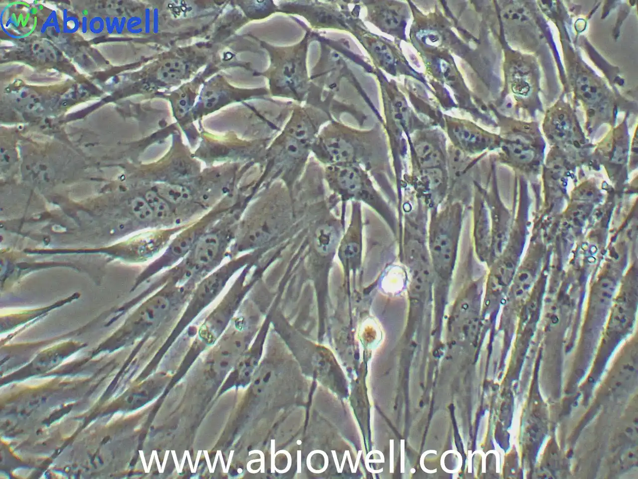
02产品简介
HL-1(小鼠心肌细胞)(STR鉴定:规格 1×10⁶cells。
03产品介绍
认真做好每一个抗体, 每一个抗体都经过反复验证!
此HL-1心肌细胞系是一个永生的老鼠心肌细胞细胞系能够自发地不断分裂和合同同时保持心脏表型分化。HL-1可以连续通道没有失去心脏肌细胞表型分化,包括形态学、生物化学和电生理特性。HL-1用于各种模型系统解决问题的心脏生物在细胞和分子水平。
1) 来源:小鼠,雌性,心肌
2) 形态:成纤维细胞样,贴壁生长
空气,95%;二氧化碳,5%
1) 收到细胞后,活细胞首先观察培养瓶是否完好,培养液是否漏液,培养基是否浑浊;冻存细胞是否干冰已挥发完,冻存管盖是否脱落,破碎,若有这类情况,请务必拍照记录,并于收货24h内与我们联系。
复苏的细胞:如果是T-25培养瓶活细胞,收到后请用75%的酒精对培养瓶表面进行消毒处理,然后转入培养箱中静置2~3h后再进行后续处理。
备注:运输用的培养基不宜再次用来培养细胞,请按照 新配置完全培养基来培养细胞。
冻存细胞:如果是干冰运输的冻存细胞,收到后请立即转入液氮存储或者短暂(24h)放置-80度冰箱保存,或者直接进行细胞复苏。
细胞复苏、传代及冻存流程参考
1) 配制完全培养基:基础培养基+胎牛血清+双抗(特殊培养基特殊配置)
2) 细胞复苏:取5ml完全培养基于15ml离心管中,37℃水浴锅预热,从液氮管(或者-80度冰箱)中快速取出冻存的细胞,放入37℃水浴锅中,摇晃使快速化冻(1min左右),然后将化冻的细胞和预热的培养基,移入超净工作台中,化冻的细胞加入到含预热培养基的15ml离心管中,1000rpm离心5min
3) 吸弃上清,得到细胞沉淀,用2ml完全培养基轻轻重悬细胞,加入到T25培养瓶中,做好标记,放入37℃,5%CO2饱和适度培养箱中培养(培养皿复苏效果更好)
4) 24h后,观察细胞贴壁情况(未贴壁的即为死细胞--针对贴壁细胞),吸弃旧培养基,加入新鲜的预热(室温或37℃)的完全培养基,继续培养。
1) 待细胞生长到80%-90%汇合度时,吸弃旧的培养基,加入1ml无菌PBS润洗一次,以去除残余的培养基及血清(血清含有胰酶的抑制因子),然后加入1ml 0.25%胰酶,37℃培养箱中消化(1~2min左右,不同细胞消化时间不同),取出细胞,镜下观察细胞至细胞皱缩变圆
2) 加入1ml完全培养基(含FBS)终止消化,轻轻拍打,使细胞脱落下来成单个细胞悬液,收集细胞于15ml无菌离心管中,1000rpm,离心5min
3) 收集细胞沉淀,完全培养基重悬,一分为二(可根据细胞生长速度调整比例),分别加入到2个新的培养瓶中,做好标记,放入培养箱中培养。
1) 按照细胞传代方法,在超净工作台内消化收集细胞沉淀,取少量细胞用于计数
2) 用预冷的1ml冻存液(90%完全培养基+10%DMSO)或者无血清细胞冻存液重悬细胞,加入到1.2ml冻存管中,密度为1*106个/ml。
3) 放入程序冻存盒,-80℃过夜后,转入液氮长期保存。
Background Sepsis-induced cardiomyopathy (SICM) is a critical cardiovascular complication characterized by cardiac dysfunction and high mortality. The molecular mechanisms that underlie SICM remain elusive, and effective therapies are limited. Results Here, we report a pivotal role for adenosine deaminases acting on RNA-1 (ADAR1) in modulating macrophage polarization and exosome-mediated intercellular communication, which ameliorates myocardial damage in SICM. We determined that ADAR1 overexpression in macrophages promotes an anti-inflammatory M2 phenotype, reduces myocardial inflammation, and inhibits cardiomyocyte apoptosis in a murine model of sepsis. Mechanistically, ADAR1 regulates the level of microRNA-122 (miR-122) in macrophage-derived exosomes. Exosomal miR-122 targets X-linked inhibitor of apoptosis protein (XIAP), modulating cardiomyocyte survival. Conclusions Our study reveals a novel ADAR1-miR-122-XIAP axis in macrophage exosomes that protects against sepsis-induced myocardial injury, offering a potential disease modulation strategy for SICM. Graphical abstract The alternative text for this image may have been generated using AI.
Authors:Wang Linxiao, Liu Shanshou, Wang Xianqi, Wu Huirong, Zhao Xiaojun, Wu Dan, Wang Qianmei, Wang Peiwen, Xu Yanan, Shen Kuo, Yuan Juzheng, Li Junjie
Sleep disorders increase the risk and mortality of heart disease, but the brain-heart interaction has not yet been fully elucidated. Cuproptosis is a copper-dependent type of cell death activated by the excessive accumulation of intracellular copper. Here, we showed that 16 weeks of sleep fragmentation (SF) resulted in elevated copper levels in the male mouse heart and exacerbated myocardial ischemia–reperfusion injury with increased myocardial cuproptosis and apoptosis. Mechanistically, we found that SF promotes sympathetic overactivity, increases the germination of myocardial sympathetic nerve terminals, and increases the level of norepinephrine in cardiac tissue, thereby inhibits VPS35 expression and leads to impaired ATP7A related copper transport and copper overload in cardiomyocytes. Copper overload further leads to exacerbated cuproptosis and apoptosis, and these effects can be rescued by excision of the sympathetic nerve or administration of copper chelating agent. Our study elucidates one of the molecular mechanisms by which sleep disorders aggravate myocardial injury and suggests possible targets for intervention.
Authors:Chen Na, Guo Lizhe, Wang Lu, Dai Sisi, Zhu Xiaocheng, Wang E.
Monitoring the morphological and biochemical information of neurons and glial cells at high temporal resolution in three-dimensional (3D) volumes of in vivo is pivotal for understanding their structure and function, and quantifying the brain microenvironment. Conventional two-photon fluorescence lifetime volumetric imaging speed faces the acquisition speed challenges of slow serial focal tomographic scanning, complex post-processing procedures for lifetime images, and inherent trade-offs among contrast, signal-to-noise ratio, and speed. This study presents a two-photon fluorescence lifetime volumetric projection microscopy using an axially elongated Bessel focus and instant frequency-domain fluorescence lifetime technique, and integrating with a convolutional network to enhance the imaging speed for in vivo neurodynamics mapping. The proposed method is validated by monitoring intracellular Ca 2+ concentration throughout whole volume, tracking microglia movement and microenvironmental changes following thermal injury in the zebrafish brain, analyzing structural and functional variations of gap junctions in astrocyte networks, and measuring the Ca 2+ concentration in neurons in mouse brains. This innovative methodology enables quantitative in vivo visualization of neurodynamics and the cellular processes and interactions in the brain.
Authors:Yanping Li, Xiangcong Xu, Chao Zhang, Xuefeng Sun, Sisi Zhou, Xuan Li, Jiaqing Guo, Rui Hu, Junle Qu, Liwei Liu Authors:Tao Yang, Pengcui Wu, Luping Jiang, Ran Chen, Qiao Jin, Guohong Ye APPLIED BIOCHEMISTRY AND BIOTECHNOLOGY IF:3.5 Authors:Liao Dongshan, Wang Zhisheng, Tian Gengyan, Wang Honghao, Fang Guanhua Authors:Wang Ting, Xiao Helong, Yang Meijian, Liu Jing, Yang Yang Authors:Zeng Xianghui, Wang Dian, Yang Hao, Li Canqun, Xiong Ting, Lu Ting, Dai Lu, Chen Feng, Liu Zhenfang, Li Xing
Acute myocardial infarction (AMI) is a cardiovascular illness with the highest disability and mortality rates worldwide. This study aimed to estimate the mechanism of TDRG1 in myocardial damage.qRT-PCR was used to study the levels of TDRG1. After establishing hypoxia/reoxygenation (H/R) model, the inflammation was assessed by qRT-PCR, oxidation was detected by commercial kits, and apoptosis was estimated by qRT-PCR and flow cytometry. The luciferase intensity and RNA immunoprecipitation assay were detected for the identification of target relationship. The functional enrichment was unveiled by GO and Kyoto Encyclopedia of Genes and Genomes (KEGG). The protein interaction was conducted for screening key genes.The expression of TDRG1 was elevated and negatively correlated with miR-330-5p in the serum AMI patients. TDRG1/miR-330-5p axis regulated inflammation, oxidation, and viability and apoptosis of HL-1 cells induced by H/R. GO and KEGG analyses indicate that 76 overlapping targets of miR-330-5p were primarily involved in focal adhesion, calmodulin binding, and ErbB and Rap1 signaling pathways. MAPK1 was the top key gene and was a target gene of miR-330-5p.TDRG1/miR-330-5p axis could participate in the regulation of apoptosis and inflammation of H/R-induced cardiomyocytes.
Authors:Wenjing Wang, Yanhui Ni, Guangyun Cao, Xinning Li 细胞描述: GH3细胞系是由Tashjian AH等在1965年7月从一只7月龄的雌性Wistar-Furth大鼠的垂体肿瘤中分离建立的。GH3细胞系不是直接来源于GH1细胞系的克隆,而是从原代培养的GH1细胞在大鼠身上传代两次形成的肿瘤中建立的。上皮样的GH3细胞比GH1分泌更高水平的生长激素,也可产生细胞描述: 胶质细胞株C6是由Benda等用N-nitrosomethylurea诱导的大鼠胶质瘤克隆,并经过一系列的体外培养和动物传代交替后建成的。当细胞从低密度生长到满瓶时,S-100产量增加10倍。 细胞特性: 1)来源:大鼠,胶质瘤 2)形态:成纤维细胞样,贴壁生长 3) 6 cells 细胞描述: 克隆Neuro-2A是由R·J·Klebe和F·H·Ruddle经A株白鼠的自生肿瘤建立。该细胞产生大量微管蛋白,此蛋白在神经细胞中起应答轴浆流动的收缩系统作用。 细胞特性: 1)来源:脑神经母细胞瘤 2)形态:阿米巴样干细胞,贴壁生长 3) 6 cells 4)培养条件: MEM+
细胞描述: 此AML12(α小鼠肝脏12)细胞系由来自针对人TGFα的转基因小鼠(CD1菌株,品系MT42)的肝细胞建立。通过电子显微镜观察,这些细胞表现出典型的肝细胞特征,例如过氧化物酶体和胆小管样结构。AML12细胞保留表达血清(白蛋白,α1抗胰蛋白酶和转铁蛋白)和间隙连细胞描述: OVCAR-3细胞由T·C·Hamilton在1982年建系。取于患进行性卵巢腺癌病人的恶性腹水。NIH-OVCAR-3是研究卵巢癌药物抗性的一个合适的模型系统且由于存在激素受体,这对于激素治疗的评估或许是有用的。 细胞特性: 1)来源: 卵巢 2)形态:上皮细胞样,贴壁生长 3)规格细胞描述: 该细胞是从一名13岁的白人女性的骨肉瘤组织中分离建立的,表现为扁平形态、低饱和密度、软琼脂中的低克隆形成率和对化合物及病毒感染的敏感性。 细胞特性: 1)来源:骨 2)形态:上皮细胞样,贴壁生长 3) 6 cells 4)培养条件: MEM+10%FBS+1%NEAA+1 细胞描述: 该细胞系于1982年建立,从一位56岁白种男性的病变舌中间部分取得,该细胞具高密度颗粒状胞浆。 细胞特性: 1)来源:舌 2)形态:上皮细胞样,贴壁生长 3) 6 cells 4)培养条件: DMEM+10%FBS+1%P/S 空气,95%;二氧化碳,5% 37℃ 细胞接收后的处理: 1)收
细胞描述: HEK-293T细胞是293T(293tsA1609neo)细胞系(ATCC CRL-11268)的衍生物。细胞持续表达SV40抗原,该细胞常用于转染实验,转染效率较高。(转染一般以50%密度铺板,待细胞刚刚贴到皿壁上后(一般8-10小时),即可进行转染操作)。 细胞特性: 1)来源: 人胚胎肾脏 2)形这株细胞于1988年七月建株,组织提供者是一位非吸烟人士。
该人胰腺癌细胞PANC-1来源于一名56岁白人男性。1 U/ml 左旋天冬酰胺酶(L-asparaginase)可抑制其生长。此细胞可以在琼脂糖上生长。其倍增时间为52小时,G6PD活性处于低泳动性的B型。染色体研究表明模式数目为63,包括3个独特标记的染色体和1个小环状染色体。 以下二点证明它的恶性:(1)在软琼脂上和单层成纤维细胞上的快速生长,(2)注入无胸腺裸鼠后形成日益增长的成长型未分化癌。
相关资料下载:说明书(.pdf)(点击从官网下载)
04完整参数表
参数由厂商资料整理,采购前请以最终报价单与技术协议为准。
05荣誉与资质
该品牌暂未上传荣誉证书与资质文件。以下是平台可核验的公开信息,未上传项如实标注,不做推断。
- 营业执照未上传
- 品牌授权未上传
- 产品检测报告未上传
- 会员身份
- 收费会员
- 经营身份
- 原厂
- 成立年份
- 2016 年
- 所在地
- 湖南
- 收录条目
- 498 个
- 平台核验
- 平台代管
06品牌与供应商
公司产品涵盖抗体、细胞、培养基、质粒载体等多个领域,致力于为生命科学研究提供可靠的工具与解决方案。自创立以来,艾碧维始终秉持“求实、诚挚、勤勉、克己”的发展理念,坚持以自主研发与持续创新为核心动力。公司拥有百余人的团队,其中硕士及以上学历生物医学专业人才占比超过30%,包括博士4人,构建起一支结构合理、专业扎实的研发与服务队伍。通过设立研发部、生产部、市场与营销部、财务部等多功能部门,公司实现了从产品开发到市场推广的全流程管控。艾碧维不仅敢于质疑现状,积极为生命科学家突破技术壁垒提供支撑,更注重与客户的深度互动与合作,通过持续收集反馈、开展研发协作,提前预见并解决潜在问题。未来,公司将继续秉持“助力科研创新,让实验更简单”的使命,一步一个脚印地打造具有国际领先水平的技术研发与产业化平台,为全球生命科学领域的进步持续赋能。注意: 1.本公司产品仅供科研使用。
品牌资料由厂商公开信息与授权渠道核对整理;条目参数与价格以最终报价单为准。
07同类条目
同一子类内的条目才具备可比性。跨子类比较参数通常没有意义。
08同品牌其他条目
艾碧维 在本目录共 498 个条目,除本条外还有:
科研在线(TeleVM). HL-1(小鼠心肌细胞)(STR鉴定)(AW-CNM080)[EB/OL]. 产品目录, 2026-10-31. https://www.suprotek.cn/products/item/aw-cnm080.html.
TeleVM. (2026, October 31). HL-1(小鼠心肌细胞)(STR鉴定)(AW-CNM080). 产品目录. https://www.suprotek.cn/products/item/aw-cnm080.html
@misc{biocsp_item_aw_cnm080,
  title        = {HL-1(小鼠心肌细胞)(STR鉴定)(AW-CNM080)},
  author       = {{科研在线}},
  year         = {2026},
  howpublished = {产品目录},
  note         = {更新于 2026-10-31,产品目录},
  url          = {https://www.suprotek.cn/products/item/aw-cnm080.html}
}
GB/T 7714 用于中文论文与报告;APA 7 用于英文期刊;BibTeX 可直接导入 Zotero、EndNote 或 LaTeX。
许可:CC BY-NC 4.0 · 转载注明来源