
iF647 Anti-Mouse CD69 Antibody
英诺赛图(InnoCyto)的 iF647 Anti-Mouse CD69 Antibody(克隆 H1.2F3) 为Armenian Hamster IgG 单克隆抗体,靶向 CD69,iF647 标记(633 nm 红色激光激发),适用于流式细胞术。提供 25 μg、100 μg 规格,目录价 ¥1,155 起,仅供科研使用(RUO)。
01产品图片
02产品简介
英诺赛图(InnoCyto)的 iF647 Anti-Mouse CD69 Antibody(克隆 H1.2F3) 为Armenian Hamster IgG 单克隆抗体,靶向 CD69,iF647 标记(633 nm 红色激光激发),适用于流式细胞术。提供 25 μg、100 μg 规格,目录价 ¥1,155 起,仅供科研使用(RUO)。
03产品介绍
规格与货号
204507 | 25 μg | ¥1,155
204508 | 100 μg | ¥2,645
Product Details
Regulatory Status|RUO
Protein Concentration|Supplied at a lot-specific concentration.
Storage & Handling|The antibody solution should be stored undiluted between 2°C and 8°C, and protected from prolonged exposure to light. Do not freeze.
Recommended Usage|For flow cytometric staining, it is recommended to use less than 0.3 μg of this reagent per 0.5-1.0 million cells in a 100 μL volume. Optimal reagent performance should be determined by titration for each specific application. iF647 has an excitation max at 656 nm and an emission max at 670 nm.
See All Formats|Clone H1.2F3
Background Information
CD69 is a type II transmembrane C-type lectin receptor that is best known as one of the earliest inducible markers of immune cell activation. It is rapidly upregulated on the surface of T cells, B cells, natural killer (NK) cells, dendritic cells, and other leukocytes following antigen receptor engagement, cytokine stimulation, or inflammatory signaling. While CD69 has long been used as a phenotypic activation marker, it is now recognized as an active regulator of immune cell localization and function.
Structurally, CD69 is expressed as a disulfide-linked homodimer. Each subunit contains a short N-terminal cytoplasmic tail, a single-pass transmembrane domain, and a C-terminal extracellular C-type lectin-like domain. Despite its classification within the C-type lectin family, CD69 does not require calcium for ligand binding. The cytoplasmic region includes signaling motifs that allow CD69 to associate with intracellular adaptor proteins and influence downstream signaling pathways, contributing to modulation of immune responses.
The most well-characterized ligand for CD69 is sphingosine-1-phosphate receptor 1 (S1PR1), a G protein–coupled receptor that controls lymphocyte egress from lymphoid organs into the circulation. CD69 physically associates with S1PR1, promoting its internalization and functional inhibition. This interaction restrains the migration of activated lymphocytes, leading to their retention within lymphoid tissues or sites of inflammation. Through this mechanism, CD69 plays a key role in the establishment and maintenance of tissue-resident memory T cells, which provide rapid, localized immune protection.
CD69 is involved in a broad range of disease processes. In infectious disease, CD69 expression marks recently activated effector cells and tissue-resident populations that contribute to protective immunity. In autoimmune and chronic inflammatory diseases, persistent CD69 expression can promote the accumulation of pathogenic lymphocytes in affected tissues, potentially exacerbating disease. Conversely, CD69 has also been shown to exert immunoregulatory effects by dampening excessive immune activation, highlighting its dual, context-dependent role. Elevated CD69 expression is commonly observed on tumor-infiltrating lymphocytes and may reflect ongoing immune activation or tissue residency within the tumor microenvironment.
From a therapeutic perspective, CD69 is primarily used as a biomarker to monitor immune activation, lymphocyte trafficking, and tissue residency in research and clinical settings. Although CD69 itself is not a major direct drug target, its functional interaction with S1PR1 has informed the development of therapies that modulate lymphocyte migration, such as sphingosine-1-phosphate receptor modulators used in autoimmune disease. Ongoing studies of CD69 biology continue to inform immunotherapeutic strategies aimed at enhancing protective immunity or limiting pathological inflammation.
Data Sheets
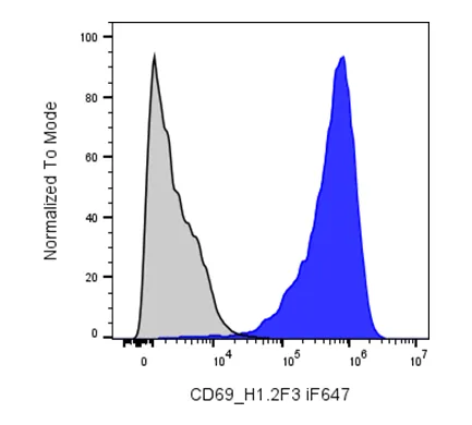
BALB/c Mouse splenocytes stimulated with Anti-Mouse CD3/CD28 for 2-days were stained with i F647 Anti- Mouse CD69 clone H1.2F3 (color-filled histogram) or an isotype control (gray histogram).
04完整参数表
参数由厂商资料整理,采购前请以最终报价单与技术协议为准。
05荣誉与资质
该品牌暂未上传荣誉证书与资质文件。以下是平台可核验的公开信息,未上传项如实标注,不做推断。
- 营业执照未上传
- 品牌授权未上传
- 产品检测报告未上传
- 会员身份
- 收费会员
- 经营身份
- 原厂
- 成立年份
- 2020 年
- 所在地
- 上海
- 收录条目
- 1865 个
- 平台核验
- 平台代管
06品牌与供应商
InnoCyto(上海英诺赛图生物科技有限公司)致力于通过开发高质量细胞分析试剂,加速科学发现进程。公司为研究人员提供细胞层面探索所需的工具,推动免疫学、肿瘤学等多领域的研究创新。 核心团队拥有丰富的行业经验,曾参与全球头部生命科学公司的标杆项目搭建,深刻理解工业界与学术研究的前沿需求。试剂从设计之初即以精确性、一致性、高性能为核心标准,让科学家能把精力集中在最核心的研究工作上。 公司将深厚的科学积累与对品质的严格承诺相结合,力争成为全球研究人员信赖的合作伙伴。产品线覆盖经验证的一抗与二抗、同型对照、生物类似药抗体、重组蛋白、细胞因子、免疫检测试剂盒及配套试剂,并提供重组蛋白/抗体表达与标记偶联服务。
品牌资料由厂商公开信息与授权渠道核对整理;条目参数与价格以最终报价单为准。
07同类条目
同一子类内的条目才具备可比性。跨子类比较参数通常没有意义。
08同品牌其他条目
上海英诺赛图 在本目录共 1865 个条目,除本条外还有:
科研在线(TeleVM). iF647 Anti-Mouse CD69 Antibody(204507)[EB/OL]. 产品目录, 2026-11-12. https://www.suprotek.cn/products/item/if647-anti-mouse-cd69-antibody.html.
TeleVM. (2026, November 12). iF647 Anti-Mouse CD69 Antibody(204507). 产品目录. https://www.suprotek.cn/products/item/if647-anti-mouse-cd69-antibody.html
@misc{biocsp_item_if647_anti_mouse_cd69_antibody,
  title        = {iF647 Anti-Mouse CD69 Antibody(204507)},
  author       = {{科研在线}},
  year         = {2026},
  howpublished = {产品目录},
  note         = {更新于 2026-11-12,产品目录},
  url          = {https://www.suprotek.cn/products/item/if647-anti-mouse-cd69-antibody.html}
}
GB/T 7714 用于中文论文与报告;APA 7 用于英文期刊;BibTeX 可直接导入 Zotero、EndNote 或 LaTeX。
许可:CC BY-NC 4.0 · 转载注明来源